|
Case Report
Nephrogenic adenoma in a female urethral diverticulum: Clinical presentation and surgical management
1 Division of Urogynecology and Reconstructive Pelvic Surgery, Department of Obstetrics and Gynecology, University of Iowa Hospitals and Clinics, Iowa City, IA 52242, USA
2 Medical Student, Roy J. and Lucille A. Carver College of Medicine, The University of Iowa, Iowa City, IA 52242, USA
3 Department of Pathology, University of Iowa Hospitals and Clinics, Iowa City, IA 52242, USA
4 Department of Urology, 200 Hawkins Drive, University of Iowa Hospitals and Clinics, Iowa City, IA 52242, USA
Address correspondence to:
Leanne Brechtel
200 Hawkins Ave, Iowa City, IA 52242,
USA
Message to Corresponding Author
Article ID: 100068Z15LB2026
Access full text article on other devices

Access PDF of article on other devices


How to cite this article
Brechtel L, Venkatesh V, Christensen N, Stauffer S, Vollstedt A. Nephrogenic adenoma in a female urethral diverticulum: Clinical presentation and surgical management. J Case Rep Images Urol 2026;11(2):7–11.ABSTRACT
Introduction: Nephrogenic adenoma is a benign lesion of the urinary tract that is rarely found within a urethral diverticulum. The nonspecific symptoms of urethral diverticula can often lead to delayed or missed diagnosis, complicating management.
Case Report: We present the case of a woman who presented with vaginal discomfort and was ultimately found to have a periurethral mass. Pelvic magnetic resonance imaging (MRI) confirmed the presence of a periurethral mass. Definitive surgical excision and histopathological analysis confirmed the presence of nephrogenic adenoma within the urethral diverticulum. Postoperatively, the patient did well with near resolution of her symptoms.
Conclusion: This case highlights the importance of a broad differential diagnosis, the utility of advanced imaging, and relying on histopathology to distinguish between benign and malignant lesions. For nephrogenic adenoma, surgical excision remains the optimal treatment, often offering excellent outcomes.
Keywords: Histopathology, Nephrogenic adenoma, Surgical technique, Urethral diverticulum, Vaginal surgery
Introduction
Nephrogenic adenoma is a rare benign neoplasm that may occur throughout the urinary tract, most frequently within the bladder. Occurrence within a urethral diverticulum is uncommon, with only a small number of cases described in the literature [1],[2]. In female patients, approximately one-fourth of urethral nephrogenic adenoma cases are associated with urethral diverticulum [3]. Urethral diverticula themselves are uncommon in women and are likely underdiagnosed due to their diverse etiology and symptomology of the lower urinary tract. The classic triad of dysuria, dyspareunia, and post-void dribbling is only present in a minority of patients, and many present with varying symptoms. In the context of nephrogenic adenoma arising within a urethral diverticulum, the clinical presentation does not differ significantly from that of the urethral diverticulum alone. In most cases, they are discovered incidentally during excision of the diverticulum. However, given the risk of neoplastic lesions, including malignancy, careful clinical evaluation and imaging are warranted. Such lesions present challenges that stem from their rarity, histologic overlap with malignant neoplasms, and incidental discovery after surgical excision [4],[5]. We present a unique case of a patient with a periurethral mass treated initially with topical therapy, aborted surgical cyst removal after intraoperative discovery of urethral diverticulum, and pathology of nephrogenic adenoma after undergoing diverticulectomy, resulting in significant symptom improvement postoperatively.
Case Report
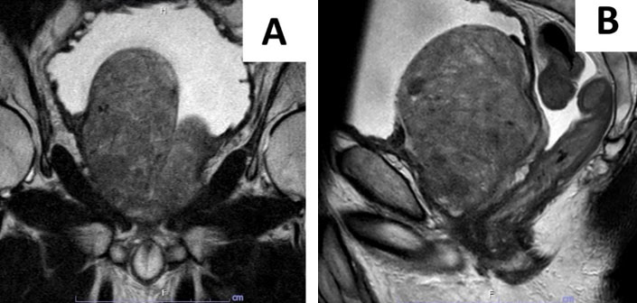
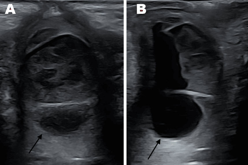
The patient is a 43-year-old female with a medical history significant hypothyroidism on levothyroxine, seborrheic dermatitis and endometriosis. Her surgical history includes a unilateral salpingo-oophorectomy in 2016 for endometrioma and a breast biopsy. She initially presented to her primary care provider (PCP) with complaints of vaginal soreness, swelling, and pruritus. Suspecting a yeast infection, she had tried over-the-counter miconazole suppositories without relief. At that time, she denied abnormal discharge and malodor. Physical examination in the clinic documented mild vulvar swelling without other abnormalities, and a vaginal swab for bacterial vaginosis and yeast testing was obtained. She was prescribed a three-day course of vaginal terconazole cream due to an allergy to oral fluconazole. Although her symptoms initially improved, she reported recurrent swelling and discomfort after completing treatment. She returned to her primary care physician the following month with persistent swelling and discomfort, along with increased pressure during urination and a slow urinary stream. On physical exam, she was noted to have slight swelling over the anterior vaginal wall. She had a vaginitis panel, which was negative, but despite this, a repeat course of terconazole was prescribed. She was then referred to a gynecologist for further evaluation. Upon presentation to the gynecologist, she reported a non-painful vaginal mass, without dysuria or abnormal discharge. Physical examination at that time revealed an approximately 3 cm smooth mass on the anterior vaginal wall, prompting suspicion of prolapse or a periurethral cyst. A trans-labial ultrasound subsequently demonstrated a 3.7 cm complex cystic structure with circumferential vascularity and no discernible communication with any adjacent pelvic structures. Based on ultrasound findings of a well-circumscribed cyst without a tract to the urethral lumen, a Skene’s gland cyst was suspected (Figure 1). She then underwent surgery for excision of the presumed Skene’s gland cyst with gynecology. Intraoperatively, compression of the mass resulted in the expression of copious gray fluid from the urethral meatus, observed around the Foley catheter. Due to concerns regarding a urethral diverticulum, the procedure was terminated, and the patient was referred to urology for further management. Fluid cultures of the cyst contents yielded no evidence of infection. At her initial urology consultation, the patient reported post-void dribbling of urine and pelvic pressure with intercourse. In the clinic, physical examination revealed a firm, periurethral mass measuring approximately 2 × 4 cm, extending along the length of urethra. To further evaluate the mass, the patient underwent a pelvic MRI. The MRI revealed a “cystic lesion in the low midline pelvis containing dependent material and closely associated with the anterior vaginal wall, with broad contact along the posterior urethra” (Figure 2). Surgical excision was therefore pursued, and the patient was taken to the operating room for transvaginal removal of the mass. The patient was placed in dorsal lithotomy position. The large periurethral cyst was again noted, spanning the entire length of the urethra, with palpation prompting purulent urine to leak from the meatus. The cloudy urine was sent for culture, and the procedure began with a cystoscopy using a 17 French rigid cystoscope. No abnormalities were seen on cystourethroscopy and no os in the urethra was initially visualized. No significant scar tissue was present. A 14 French Foley catheter was then placed. Using a LoneStar retractor for visualization, an inverted U-Flap was created in the vaginal epithelium, and the anterior vaginal wall was carefully reflected to expose the diverticulum. The periurethral fascia was dissected away from the diverticular sac, and the diverticulum was entered. To achieve better visualization of the connection to the urethra, the Foley catheter was removed and replaced with a flexible cystoscope. At this time, a posterior midline os at the 6 o’clock position was visualized and cannulated with a sensor wire, which was then seen existing the urethral defect. The defect in the urethra was closed using a running 4-0 Vicryl. The closure of the urethral was deemed to be water-tight after injecting methylene blue alongside the catheter using a 14-gauge angiocatheter. The diverticular sac tissue was excised along the urethra and sent to pathology. The wall of the urethral diverticulum was noted to appear thicker than a typical urethral diverticulum. However, no other masses or suspicious tissue were encountered. Probing with a lacrimal duct probe confirmed the absence of additional cystic components. The periurethral tissue and fascia were then closed using 3-0 Vicryl, and vaginal epithelium was closed using a 2-0 Vicryl in a non-overlapping fashion. The catheter was removed and repeat cystoscopy demonstrated efflux from bilateral ureteral orifices. A 16 French catheter was then placed and attached to a drainage bag. The patient tolerated the procedure well and was discharged the same day.
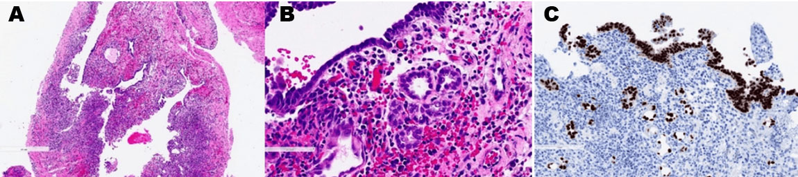
Microscopic pathologic evaluation of the excised diverticulum demonstrated a tubular and glandular architecture with a characteristic hobnail appearance, characterized by nuclei protruding into the luminal space. Immunohistochemical staining showed the lesional cells to be positive for PAX-8 and negative for carcinoembryonic antigen (CEA), WT-1, and GATA-3, with a low Ki-67 proliferative index (Figure 3). The morphologic and immunophenotypic findings were consistent with a diagnosis of nephrogenic adenoma.
She had her first follow-up appointment two weeks following her excision and at that time, she reported good wound healing and resolution of her symptoms. Urine culture from the time of her procedure indicated no evidence of infection. She had no dysuria, hematuria, or evidence of infection. After a successful trial of void, her Foley catheter was removed. She was advised to adhere to lifting restrictions, maintain pelvic rest for six weeks, and ensure adequate fluid intake. She remains under routine clinical surveillance for recurrence. Given her continued absence of symptoms, no additional intervention is planned unless new symptoms arise.

Discussion
Nephrogenic adenoma is a rare benign lesion of the urothelium that is thought to result from implantation and proliferation of renal tubular cells in areas of urothelial injury or chronic inflammation. Proposed etiologies including persistent inflammation, recurrent infection, prior surgery, trauma, catheterization, and urinary calculi. In this case, the patient’s history of pelvic surgery and endometriosis may have contributed to a chronic inflammatory environment that predisposed to lesion development.
Nephrogenic adenomas are typically found in the bladder, less often in other parts of the urinary tract, like the urethra and ureter, and are very rarely seen in the renal pelvis [5]. In this patient, the initial presentation of vaginal soreness and swelling was nonspecific and first treated as an infection with topical antifungal therapy. The eventual identification of a periurethral mass prompted surgical intervention. The intraoperative discovery of the diverticulum after initially being treated by her PCP for vaginitis and then her gynecologist for a Skene’s gland cyst is consistent with previously reported literature that shows that urethral diverticula are often misdiagnosed for over five years [6],[7]. Magnetic resonance imaging played a crucial role in delineating the lesion, consistent with prior literature emphasizing its value in diagnosing and characterizing urethral diverticula [8]. Although not obtained prior to her index surgery in this case, pelvic MRI should be completed prior to initial surgical intervention for more accurate diagnosis.
Most reported cases of nephrogenic adenoma within urethral diverticula describe incidental diagnosis following surgical excision of presumed periurethral cysts or masses, similar to our case. Histologically, nephrogenic adenoma may mimic malignancies such as urothelial carcinoma and clear-cell adenocarcinoma, necessitating full dissection and pathologic evaluation including histopathology and immunohistochemical staining for accurate differentiation [9]. The positive PAX8 staining and characteristic tubular morphology in this case supported the benign diagnosis, avoiding unnecessary oncologic treatments.
Given the absence of recurrent urinary tract infections, which are often a symptom associated with urethral diverticulum, we did not initiate prophylactic long-term antibiotics following nephrogenic adenoma resection. Although the AUA suggests consideration of antibiotic prophylaxis, the evidence remains variable [10],[11]. Surgical excision remains the primary treatment for urethral diverticulum and is typically curative, with recurrence occurring infrequently [12],[13]. The patient experienced complete resolution of symptoms following diverticulectomy, further supporting the effectiveness of surgical management.
Conclusion
Nephrogenic adenoma arising within a urethral diverticulum is an exceptionally rare condition that poses diagnostic and therapeutic challenges because of its nonspecific presentation, frequent misdiagnosis, and histologic similarity to other lesions. In this case, advanced imaging and detailed histopathologic assessment were critical in establishing the diagnosis.
Complete surgical excision of the diverticulum and associated lesion remains the foundation of treatment and is associated with favorable outcomes and low recurrence rates. This case emphasizes the importance of maintaining a broad differential diagnosis in women presenting with persistent periurethral or vaginal symptoms and highlights the value of a multidisciplinary approach incorporating imaging, pathology, and clinical evaluation to achieve accurate diagnosis and appropriate management.
REFERENCES
1.
Sidana A, Zhai QJ, Mahdy A. Nephrogenic adenoma in a urethral diverticulum. Urology 2012;80(2):e21–2. [CrossRef]
[Pubmed]

2.
Thomas AA, Rackley RR, Lee U, Goldman HB, Vasavada SP, Hansel DE. Urethral diverticula in 90 female patients: A study with emphasis on neoplastic alterations. J Urol 2008;180(6):2463–7. [CrossRef]
[Pubmed]
3.
Amin W, Parwani AV. Nephrogenic adenoma. Pathol Res Pract 2010;206(10):659–62. [CrossRef]
[Pubmed]
4.
Pincus JB, Laudano M, Leegant A, Downing K. Female urethral diverticula: Diagnosis, pathology, and surgical outcomes at an academic, urban medical center. Urology 2019;128:42–6. [CrossRef]
[Pubmed]
5.
Gordetsky J, Gennaro KH, Selph JP, Rais-Bahrami S. Nephrogenic adenoma: Clinical features, management, and diagnostic pitfalls. Urology 2016;95:29–33. [CrossRef]
[Pubmed]
6.
Wessells H, Morey A, Souter L, Rahimi L, Vanni A. Urethral stricture disease guideline amendment (2023). J Urol 2023;210(1):64–71. [CrossRef]
[Pubmed]
7.
Romanzi LJ, Groutz A, Blaivas JG. Urethral diverticulum in women: Diverse presentations resulting in diagnostic delay and mismanagement. J Urol 2000;164(2):428–33.
[Pubmed]
8.
Saidman JM, Aineseder M, Garratt J, Wang MX, Ahmed I, Elsayes KM, et al. Imaging the female urethra: US and MRI in cystic and solid pathologic conditions. Radiographics 2025;45(3):e240064. [CrossRef]
[Pubmed]
9.
López JI, Schiavo-Lena M, Corominas-Cishek A, Yagüe A, Bauleth K, Guarch R, et al. Nephrogenic adenoma of the urinary tract: Clinical, histological, and immunohistochemical characteristics. Virchows Arch 2013;463(6):819–25. [CrossRef]
[Pubmed]
10.
Lightner DJ, Wymer K, Sanchez J, Kavoussi L. Best practice statement on urologic procedures and antimicrobial prophylaxis. J Urol 2020;203(2):351–6. [CrossRef]
[Pubmed]
11.
Voss K, Peppas D. Recurrent nephrogenic adenoma: A case report of resolution after treatment with antibiotics and nonsteroidal anti-inflammatory medication. Urology 2013;82(5):1156–7. [CrossRef]
[Pubmed]
12.
Chen X, Ang X, Xu X, You J, Chen W, Huang Y, et al. Experience in conversion of symptomatic urethral diverticulum to asymptomatic status through surgery. Sci Rep 2025;15(1):7533. [CrossRef]
[Pubmed]
13.
Gujral H, Chen H, Ferzandi TR. Nephrogenic adenoma in a urethral diverticulum. Female Pelvic Med Reconstr Surg 2014;20(6):e12–4. [CrossRef]
[Pubmed]
SUPPORTING INFORMATION
Author Contributions
Leanne Brechtel - Conception of the work, Design of the work, Acquisition of data, Analysis of data, Drafting the work, Revising the work critically for important intellectual content, Final approval of the version to be published, Agree to be accountable for all aspects of the work in ensuring that questions related to the accuracy or integrity of any part of the work are appropriately investigated and resolved.
Veena Venkatesh - Conception of the work, Design of the work, Acquisition of data, Analysis of data, Drafting the work, Revising the work critically for important intellectual content, Final approval of the version to be published, Agree to be accountable for all aspects of the work in ensuring that questions related to the accuracy or integrity of any part of the work are appropriately investigated and resolved.
Nathanael Christensen - Conception of the work, Design of the work, Acquisition of data, Analysis of data, Drafting the work, Revising the work critically for important intellectual content, Final approval of the version to be published, Agree to be accountable for all aspects of the work in ensuring that questions related to the accuracy or integrity of any part of the work are appropriately investigated and resolved.
Stephanie Stauffer - Analysis of data, Drafting the work, Revising the work critically for important intellectual content, Final approval of the version to be published, Agree to be accountable for all aspects of the work in ensuring that questions related to the accuracy or integrity of any part of the work are appropriately investigated and resolved.
Annah Vollstedt - Analysis of data, Drafting the work, Revising the work critically for important intellectual content, Final approval of the version to be published, Agree to be accountable for all aspects of the work in ensuring that questions related to the accuracy or integrity of any part of the work are appropriately investigated and resolved.
Guaranter of SubmissionThe corresponding author is the guarantor of submission.
Source of SupportNone
Consent StatementWritten informed consent was obtained from the patient for publication of this article.
Data AvailabilityAll relevant data are within the paper and its Supporting Information files.
Conflict of InterestAuthors declare no conflict of interest.
Copyright© 2026 Leanne Brechtel et al. This article is distributed under the terms of Creative Commons Attribution License which permits unrestricted use, distribution and reproduction in any medium provided the original author(s) and original publisher are properly credited. Please see the copyright policy on the journal website for more information.