|
Case Report
Robot-assisted management of a complex renal mass in the second trimester of pregnancy: A case report and review of recent literature
1 Department of Urology and Paediatric Urology, University Hospital of Wuerzburg, Oberduerrbacherstr. 6, 97080 Wuerzburg, Germany
2 Department of Radiology, University Hospital of Wuerzburg, Oberduerrbacherstr. 6, 97080 Wuerzburg, Germany
3 Department of Pathology, University of Wuerzburg, Josef-Schneider-Str. 2, 97080 Wuerzburg, Germany
4 Department of Obstetrics and Gynecology, University Hospital of Wuerzburg, Josef-Schneider-Str. 4, 97080 Wuerzburg, Germany
Address correspondence to:
Vincent Scheper
Department of Urology and Paediatric Urology, University Hospital of Wuerzburg, Oberduerrbacherstr. 6, 97080 Wuerzburg,
Germany
Message to Corresponding Author
Article ID: 100069Z15VS2026
Access full text article on other devices

Access PDF of article on other devices


How to cite this article
Scheper V, Kunz A, Herzog G, Büchel J, Pecks U, Kübler H, Seitz A. Robot-assisted management of a complex renal mass in the second trimester of pregnancy: A case report and review of recent literature. J Case Rep Images Urol 2026;11(2):12–17.ABSTRACT
Introduction: Renal tumors during pregnancy are rare and present a complex challenge and the need to consider maternal and fetal safety.
Case Report: A 32-year-old woman presented with right flank pain at 24+5 weeks of gestation. Ultrasound and magnetic resonance imaging (MRI) revealed a 10 cm hypervascular renal mass extending into the collecting system, initially suspected to be malignant (renal cell carcinoma vs angiomyolipoma). Following multidisciplinary consultation (urology, radiology, and obstetrics), a robot-assisted laparoscopic right nephrectomy was performed with a low pneumoperitoneum pressure (8 mmHg) strategy. Histopathology confirmed an angiomyolipoma. The maternal and fetal courses were without complications.
Conclusion: This case shows the feasibility of robot-assisted surgery for complex renal masses during pregnancy and demonstrates the importance of a multidisciplinary team to optimize maternal and fetal outcomes.
Keywords: Angiomyolipoma (AML) in pregnancy, Renal angiomyolipoma, Renal cell carcinoma (RCC), Robot-assisted surgery
Introduction
Renal masses during pregnancy are exceedingly rare, with an estimated incidence of approximately 1 per 100,000 pregnancies [1]. Their management poses unique diagnostic and therapeutic challenges. The most common solid renal tumors include renal cell carcinoma (RCC) and angiomyolipoma (AML). Angiomyolipoma is the most common benign mesenchymal neoplasm of the kidney and is typically discovered incidentally [2]; however, in pregnancy it poses added risk of rupture due to hormonal changes following increased growth [3]. Renal cell carcinoma carries clear oncological implications and often prompts early surgical intervention [4]. Diagnostic imaging is constrained in pregnancy [limitation of computed tomography (CT), avoidance of ionizing radiation] and the balance between maternal oncologic safety and fetal wellbeing must be carefully managed [5]. In particular, AML in pregnant patients has been associated with a significant risk of spontaneous rupture or hemorrhage during gestation or labor, prompting some teams to favor early operative management when size, symptoms or imaging features suggest high risk [6],[7],[8]. In our case, the multidisciplinary decision-making hinged on the dual differentials of AML vs RCC and the high hemorrhagic potential of the lesion—thus favoring early intervention despite pregnancy.
While open surgery has historically been the standard approach, recent advances have enabled laparoscopic and robot-assisted techniques even in select pregnant patients, promising reduced morbidity and faster recovery. However, reports of robot-assisted (partial) nephrectomy during pregnancy remain exceedingly rare [9]. As an additional therapeutic option transarterial embolization has been described in literature; however, the long-term effects of fetal radiation exposure remain to be discussed [10].
Case Report
A woman in her 30s (G1P0) presented at 24+5 weeks of gestation with right-sided flank discomfort over several weeks. Her medical, surgical and family history were unremarkable; she denied prior urinary tract infections or hematuria and had no known allergies. Her body mass index (BMI) was 25.5 kg/m2 (height 176 cm, weight 79 kg). She was classified as American Society of Anesthesiologists (ASA) III.
Preoperative laboratory investigations revealed normal electrolytes, creatinine 0.62 mg/dL, urea 12.7 mg/dL, hemoglobin 11.6 g/dL, leukocytes 7.6 × 10³/μL, and C-reactive protein (CRP) 0.28 mg/dL; urinalysis was unremarkable.
A detailed imaging work-up included renal ultrasound, Doppler studies, and magnetic resonance imaging (MRI) without gadolinium enhancement. From an obstetric standpoint, the patient presented with a singleton pregnancy appropriate for gestational age, demonstrating normal fetal anatomy on ultrasound and normal uteroplacental Doppler resistance indices. Following informed consent, antenatal corticosteroid therapy with betamethasone 12 mg intramuscularly was administered to promote fetal lung maturation, as the surgical intervention was scheduled as the fetus had reached the threshold of viability.
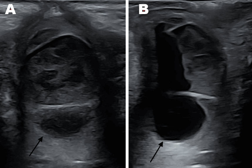
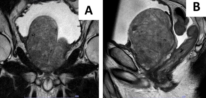
Ultrasound demonstrated right renal pelvicalyceal ectasia grade II and a ~10 cm hypervascular mass in the right kidney (Figure 1). Magnetic resonance imaging confirmed a 10 × 6.8 × 9.5 cm solid lesion located in the upper and mid pole of the right kidney, showing inhomogeneous signal intensity (predominantly T2 hypointense), central macroscopic fat components, and multiple flow voids. The mass extended into the calyceal system but showed no overt infiltrative growth or lymphadenopathy (Figure 2).
The image findings were suggestive of a RCC, but the presence of central fat and marked vascularity raised the possibility of an AML. A small cranial peritumoral hematoma was also identified.
Given the lesion’s large size (~10 cm), hypervascularity, and the risk of spontaneous rupture—particularly during labor in the case of AML—as well as the oncologic uncertainty if RCC, a multidisciplinary tumor board (urology, radiology, obstetrics, and anesthesiology) recommended early surgical intervention during the second trimester as the optimal timing.
This decision was supported by published evidence describing the increased risk of rupture for large renal AMLs during pregnancy and the safety of surgical management in the mid-gestational period. Large tumor size, pregnancy, and aneurysm or hematoma were reported as independent risk factors for bleeding. Up to 15–30% occurrence of hemorrhage has been described. Therefore, continued surveillance was considered unsafe because it would have exposed both mother and fetus to a substantial risk of hemorrhage and potentially delayed treatment of an underlying RCC.
The primary working differentials at presentation were RCC and AML. Renal cell carcinoma was considered due to the size and imaging features of the mass, including extension into the collecting system. Angiomyolipoma was likewise strong in the differential because of the presence of macroscopic fat components, hypervascularity, and the demographic context (young woman). The decision-making process favored AML as the more likely diagnosis owing to imaging features and pregnancy context, but the possibility of malignancy could not be ruled out, hence radical resection was selected.
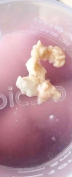
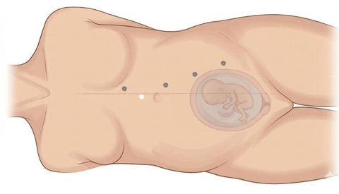
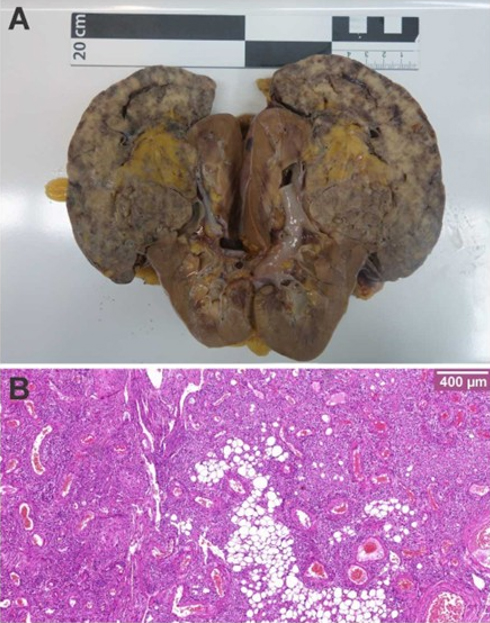
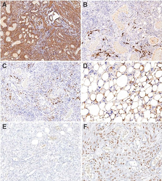
The patient underwent robotic-assisted laparoscopic right radical nephrectomy. She was positioned in 45° left lateral decubitus. A 2 cm paraumbilical mini-laparotomy was utilized for insufflation via Alexis-Laparoscopic-System and pneumoperitoneum was established at 8 mmHg CO2 pressure. Trocar placement was guided by intraoperative ultrasound to avoid uterine injury, with three robotic trocars and two assist ports (Figure 3). The transperitoneal approach was chosen due to the large tumor size and the concerns of limited space in retroperitoneoscopy. The laterocolic peritoneum was incised, the colon mobilized, ureter and gonadal vessels identified, and the renal hilum dissected. The renal artery and vein were clipped with Hem-o-lok clips. The adrenal gland was spared. The kidney was extracted via endobag through an extended trocar site. Total console time was approximately 114 minutes (skin-to-skin 8:21–10:15). Estimated blood loss was minimal; no intraoperative complications occurred. As the procedure was performed at 25+2 weeks of gestation, the surgery was conducted with obstetric and neonatology teams on standby. Pre- and postoperative cardiotocography (CTG) monitoring showed reassuring findings. Intraoperative inserted bladder catheter was removed on the first day after operation. Histopathology of the 12.5 × 5.5 × 4.9 cm specimen (558 g) revealed an 11.7 cm angiomyolipoma confined to the kidney with negative margins, no vascular invasion (Figure 4 and Figure 5). Angiomyolipomas may occasionally express hormone receptors [12]. In this case, no expression of the estrogen receptor was detected (Figure 5E). The progesterone receptor showed moderate nuclear positivity in nearly all tumor cells (Figure 5F).
Postoperative recovery was uneventful. After one night of observation on intensive care unit, the patient was transferred to a peripheral gynecological unit. At discharge, laboratory values were creatinine 0.84 mg/dL, urea 16.8 mg/dL, leukocytes 9.1 × 10³/μL, hemoglobin 10.4 g/dL. The patient was discharged on the fourth postoperative day. Urological abdominal ultrasound did not reveal any complications. Obstetric follow-ups at 28W+0, 35W+0, and 41W+0 demonstrated continued normal fetal growth, development and cephalic presentation. Additional genetic counseling was provided due to the suspicion of tuberous sclerosis as a potential underlying cause of the maternal angiomyolipoma. The patient declined invasive prenatal diagnostic testing via amniocentesis, as no fetal abnormalities or lesions were detected on ultrasound. For genetic testing for tuberous sclerosis in particular she was referred to a local doctor’s office.
The patient delivered spontaneously a healthy girl at 41+3 weeks gestation, weighing 3540 g, length 51 cm, APGAR 9/10/10, pH umbilical artery 7.22, base excess 7.2 mmol/L.


Discussion
While renal tumors in pregnancy are rare, the two most relevant entities are RCC and AML [1],[2]. Angiomyolipomas, although benign, carry a heightened risk of spontaneous rupture during pregnancy or labor due to hormonal and hemodynamic changes: pregnancy has been identified as an independent risk factor for AML hemorrhage [3],[7]. In literature, spontaneous retroperitoneal hemorrhage (Wunderlich syndrome) from AML has been documented in pregnancy, occasionally with catastrophic maternal and fetal outcomes [13]. Second, the imaging differentiation between RCC and AML can be challenging especially in pregnancy when some contrast modalities are restricted. While MRI with the smallest dose of gadolinium appears to be safe, radiological signs, such as heterogeneous signal intensities due to the varying proportions of vessels, muscle, and fat, are not specific and appear in both benign and malignant tumors [5],[11]. Third, our decision for early surgical intervention in the second trimester rested upon multiple risk factors: large size (>10 cm), high vascularity, intratumoral hematoma, and the risk of active bleeding during labor. A tumor of this size in a pregnant patient would carry a non-trivial risk of rupture; in the case of AML the literature supports early prophylactic intervention in such scenarios [6],[7],[8],[14]. In the event that the lesion represented a clinically staged T3a renal cell carcinoma (cT3a), the oncological risk would have been substantial and would equally support early intervention. Population-level data show that metastatic rates at diagnosis increase steeply with tumor size, that is, approximately 30.3% to 45.1% for tumors >7 to 10 cm [4]. Fourth, robot-assisted laparoscopic nephrectomy with low intra-abdominal pressure (8 mmHg) was feasible and safe in this gestational context. The Evidence-Based Guideline on Laparoscopy in Pregnancy published by the British Society for Gynaecological Endoscopy described safety for laparoscopy under 12 mmHg [9]. In our case, we opted for radical nephrectomy over partial nephrectomy for several reasons. First, the tumor’s anatomical complexity posed a considerable technical challenge which would have resulted in prolonged operative and ischemia times. Second, potential postoperative complications associated with partial nephrectomy, such as urine leakage (17%) and postoperative bleeding (3%) [15], were thereby avoided. This management approach is consistent with previous case reports addressing the treatment of renal masses during pregnancy [1],[6],[16]. We chose the minimal-invasive approach over open surgery in accordance to the European Association of Urology (EAU) Guidelines, which cited several studies demonstrating less morbidity and shorter hospital stays in favor for the robotic surgery [17]. Finally, the interdisciplinary management involving urology, radiology, obstetrics, and anesthesiology, together with preoperative fetal lung maturation, contributed to the favorable outcome. While in the general population renal tumors such as RCC and AML may be managed conservatively, by interventional radiology, or surgically, these options are limited in pregnant patients [17]. Ionizing radiation during angiographic procedures and the use of mTOR inhibitors carry potential teratogenic and fetotoxic risks, the latter particularly during the first and second trimesters [18]. Therefore, when active surveillance is not feasible, surgical intervention remains the mainstay of management. Potential fetal concerns during robotic surgery include reduced uteroplacental blood flow as well as hypercapnia. However, by using a low-pressure pneumoperitoneum and continuous monitoring (anesthetic: maternal arterial CO2, avoid arterial hypotension; obstetric: CTG) the risk can be minimized. Also, the risk of injury for uterus or other organs was reduced by using ultrasound for port placement [9]. This case demonstrates that a robot-assisted radical nephrectomy can be safely performed during the second trimester in a multidisciplinary setting. In line with previous reports, it underlines the importance of an individualized, team-based approach when balancing maternal and fetal safety.
Conclusion
This case highlights the expanding utility of robotic-assisted minimally invasive surgery for selected renal masses identified during pregnancy. Successful treatment requires careful assessment of the maternal and fetal risks associated with both the renal lesion and surgical intervention.
LEARNING POINTS
- Robot-assisted laparoscopic nephrectomy is feasible during the second trimester, even for large renal masses, when performed where expertise and multidisciplinary support exist.
- Differentiation between AML and RCC in pregnancy is challenging: while AML is benign, its risk of rupture justifies proactive intervention in selected cases.
- Maintaining low pneumoperitoneum pressure (≤8–12 mmHg) and adapting trocar placement for gravid anatomy helps optimize maternal and fetal safety.
- Multidisciplinary discussion and preoperative fetal preparation (including lung maturation) are key to favorable maternal–fetal outcomes.
REFERENCES
1.
Zhao Y, Yang Z, Xu W, Ji Z, Dong J. Management of renal tumors during pregnancy: Case reports. BMC Nephrol 2021;22(1):127. [CrossRef]
[Pubmed]

2.
Fujii Y, Ajima J, Oka K, Tosaka A, Takehara Y. Benign renal tumors detected among healthy adults by abdominal ultrasonography. Eur Urol 1995;27(2):124–7. [CrossRef]
[Pubmed]
3.
Boorjian SA, Sheinin Y, Crispen PL, Lohse CM, Kwon ED, Leibovich BC. Hormone receptor expression in renal angiomyolipoma: Clinicopathologic correlation. Urology 2008;72(4):927–32. [CrossRef]
[Pubmed]
4.
Monda SM, Lui HT, Pratsinis MA, Chandrasekar T, Evans CP, Dall’Era MA. The metastatic risk of renal cell carcinoma by primary tumor size and subtype. Eur Urol Open Sci 2023;52:137–44. [CrossRef]
[Pubmed]
5.
Webb JAW, Thomsen HS, Morcos SK; Members of Contrast Media Safety Committee of European Society of Urogenital Radiology (ESUR). The use of iodinated and gadolinium contrast media during pregnancy and lactation. Eur Radiol 2005;15(6):1234–40. [CrossRef]
[Pubmed]
6.
Preece P, Mees B, Norris B, Christie M, Wagner T, Dundee P. Surgical management of haemorrhaging renal angiomyolipoma in pregnancy. Int J Surg Case Rep 2015;7C:89–92. [CrossRef]
[Pubmed]
7.
Wang C, Li X, Peng L, Gou X, Fan J. An update on recent developments in rupture of renal angiomyolipoma. Medicine (Baltimore) 2018;97(16):e0497. [CrossRef]
[Pubmed]
8.
Kluiving MW, Peeters EFHI, Lely TA, van Oorschot N, de Ranitz-Greven WL. The effect of pregnancy on renal angiomyolipoma; a world of knowledge to gain, specifically in women with TSC. BMC Nephrol 2024;25(1):113. [CrossRef]
[Pubmed]
9.
Ball E, Waters N, Cooper N, Talati C, Mallick R, Rabas S, et al. Evidence-Based Guideline on Laparoscopy in Pregnancy: Commissioned by the British Society for Gynaecological Endoscopy (BSGE) Endorsed by the Royal College of Obstetricians & Gynaecologists (RCOG). Facts Views Vis Obgyn 2019;11(1):5–25.
[Pubmed]
10.
Al-Falahi A, Al-Sayyadi M, Al-Ozair B, Alwah A, Ahmed F. Wunderlich syndrome caused by spontaneous rupture of renal angiomyolipoma in the third trimester of pregnancy: Endovascular management with fetal preservation—A case report and literature review. Clin Case Rep 2025;13(2):e70183. [CrossRef]
[Pubmed]
11.
Labra A, Schiappacasse G, Constenla D, Cristi J. Renal angiomyolipomas: Typical and atypical features on computed tomography and magnetic resonance imaging. World J Radiol 2025;17(2):104282. [CrossRef]
[Pubmed]
12.
Patil PA, McKenney JK, Trpkov K, Hes O, Montironi R, Scarpelli M, et al. Renal leiomyoma: A contemporary multi-institution study of an infrequent and frequently misclassified neoplasm. Am J Surg Pathol 2015;39(3):349–56. [CrossRef]
[Pubmed]
13.
Kontos S, Politis V, Fokitis I, Lefakis G, Koritsiadis G, Simaioforidis V, et al. Rapture of renal angiomyolipoma during pregnancy: A case report. Cases J 2008;1(1):245. [CrossRef]
[Pubmed]
14.
Yamakado K, Tanaka N, Nakagawa T, Kobayashi S, Yanagawa M, Takeda K. Renal angiomyolipoma: Relationships between tumor size, aneurysm formation, and rupture. Radiology 2002;225(1):78–82. [CrossRef]
[Pubmed]
15.
Breau RH, Crispen PL, Jimenez RE, Lohse CM, Blute ML, Leibovich BC. Outcome of stage T2 or greater renal cell cancer treated with partial nephrectomy. J Urol 2010;183(3):903–8. [CrossRef]
[Pubmed]
16.
Zhang Y, Zhao T, Li Z, Jin X, Wang Z, Shen H. Retroperitoneoscopic robot-assisted laparoscopic partial nephrectomy during the second trimester of pregnancy: A case report and literature review. Int J Surg Case Rep 2025;133:111483. [CrossRef]
[Pubmed]
17.
Bex A, Ghanem YA, Albiges L, Bonn S, Campi R, Capitanio U, et al. European Association of Urology Guidelines on renal cell carcinoma: The 2025 update. Eur Urol 2025;87(6):683–96. [CrossRef]
[Pubmed]
18.
Ponticelli C, Moroni G. Fetal toxicity of immunosuppressive drugs in pregnancy. J Clin Med 2018;7(12):552. [CrossRef]
[Pubmed]
SUPPORTING INFORMATION
Acknowledgments
Declaration of generative AI and AI-assisted technologies in the manuscript preparation process
During the preparation of this work, the authors used Google Gemini in order to create Figure 4. After using this tool, the authors reviewed and edited the content as needed and take full responsibility for the content of the published article.
Vincent Scheper - Conception of the work, Design of the work, Acquisition of data, Analysis of data, Drafting the work, Final approval of the version to be published, Agree to be accountable for all aspects of the work in ensuring that questions related to the accuracy or integrity of any part of the work are appropriately investigated and resolved.
Andreas Kunz - Acquisition of data, Analysis of data, Revising the work critically for important intellectual content, Final approval of the version to be published, Agree to be accountable for all aspects of the work in ensuring that questions related to the accuracy or integrity of any part of the work are appropriately investigated and resolved.
Gloria Herzog - Acquisition of data, Analysis of data, Drafting the work, Final approval of the version to be published, Agree to be accountable for all aspects of the work in ensuring that questions related to the accuracy or integrity of any part of the work are appropriately investigated and resolved.
Johanna Büchel - Acquisition of data, Analysis of data, Drafting the work, Final approval of the version to be published, Agree to be accountable for all aspects of the work in ensuring that questions related to the accuracy or integrity of any part of the work are appropriately investigated and resolved.
Ulrich Pecks - Revising the work critically for important intellectual content, Final approval of the version to be published, Agree to be accountable for all aspects of the work in ensuring that questions related to the accuracy or integrity of any part of the work are appropriately investigated and resolved.
Hubert Kübler - Conception of the work, Design of the work, Revising the work critically for important intellectual content, Final approval of the version to be published, Agree to be accountable for all aspects of the work in ensuring that questions related to the accuracy or integrity of any part of the work are appropriately investigated and resolved.
Anna Seitz - Conception of the work, Design of the work, Revising the work critically for important intellectual content, Final approval of the version to be published, Agree to be accountable for all aspects of the work in ensuring that questions related to the accuracy or integrity of any part of the work are appropriately investigated and resolved.
Guaranter of SubmissionThe corresponding author is the guarantor of submission.
Source of SupportNone
Consent StatementWritten informed consent was obtained from the patient for publication of this article.
Data AvailabilityAll relevant data are within the paper and its Supporting Information files.
Conflict of InterestAuthors declare no conflict of interest.
Copyright© 2026 Vincent Scheper et al. This article is distributed under the terms of Creative Commons Attribution License which permits unrestricted use, distribution and reproduction in any medium provided the original author(s) and original publisher are properly credited. Please see the copyright policy on the journal website for more information.