|
Case Report
Paraganglioma in paratesticular: A rare case report
1 Urology Department, King Fahad Hospital Hofuf, AlAhsa Health Cluster, Saudi Arabia
Address correspondence to:
Ahmed Mousa Almuhanna
Urology Department, King Fahad Hospital Hofuf, AlAhsa Health Cluster,
Message to Corresponding Author
Article ID: 100043Z15AA2024
Access full text article on other devices

Access PDF of article on other devices

How to cite this article
Almuhanna AM, Alghorairy B, Alessawi TH, Albagshi SS, Alhazeem A, AlModhi HM. Paraganglioma in paratesticular: A rare case report. J Case Rep Images Urol 2024;9(1):18–21.ABSTRACT
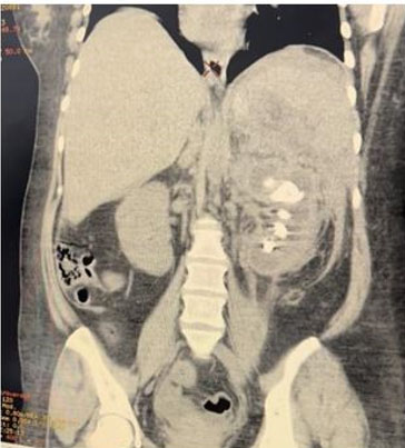
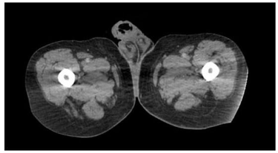
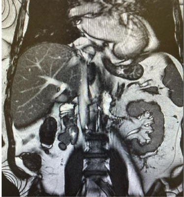
Paraganglioma at a paratesticular location is extremely rare. We report a 58-year-old Saudi male presented with two years history of right painless scrotal mass. On physical examination the scrotum revealed a right-sided non-tender mass not attached to right testis. Normal tumor markers of testicular tumor. Ultrasonography revealed a well-defined, homogeneous, hyperechoic lesion measuring approximately 2 cm in the right extratesticular region. Magnetic resonance imaging (MRI) with intravenous (IV) gadolinium contrast for abdominal and pelvis showed right extratesticular soft tissue mass not separable from the spermatic cord and there was no distant metastasis. The patient underwent exploratory excision of the mass with preservation of cord and testis. Histopathology showed paratesticular paraganglioma.
Keywords: Paraganglioma, Testicular, Tumor
SUPPORTING INFORMATION
Author Contributions
Ahmed Mousa Almuhanna - Conception of the work, Design of the work, Revising the work critically for important intellectual content, Final approval of the version to be published, Agree to be accountable for all aspects of the work in ensuring that questions related to the accuracy or integrity of any part of the work are appropriately investigated and resolved.
Basim Alghorairy - Conception of the work, Design of the work, Analysis of data, Revising the work critically for important intellectual content, Final approval of the version to be published, Agree to be accountable for all aspects of the work in ensuring that questions related to the accuracy or integrity of any part of the work are appropriately investigated and resolved.
Turki H Alessawi - Final approval of the version to be published, Agree to be accountable for all aspects of the work in ensuring that questions related to the accuracy or integrity of any part of the work are appropriately investigated and resolved.
Sara Sameer Albagshi - Drafting the work, Revising the work critically for important intellectual content, Final approval of the version to be published, Agree to be accountable for all aspects of the work in ensuring that questions related to the accuracy or integrity of any part of the work are appropriately investigated and resolved.
Abdulrahman Alhazeem - Analysis of data, Revising the work critically for important intellectual content, Final approval of the version to be published, Agree to be accountable for all aspects of the work in ensuring that questions related to the accuracy or integrity of any part of the work are appropriately investigated and resolved.
Hussain M AlModhi - Acquisition of data, Drafting the work, Final approval of the version to be published, Agree to be accountable for all aspects of the work in ensuring that questions related to the accuracy or integrity of any part of the work are appropriately investigated and resolved.
Guaranter of SubmissionThe corresponding author is the guarantor of submission.
Source of SupportNone
Consent StatementWritten informed consent was obtained from the patient for publication of this article.
Data AvailabilityAll relevant data are within the paper and its Supporting Information files.
Conflict of InterestAuthors declare no conflict of interest.
Copyright© 2024 Ahmed Mousa Almuhanna et al. This article is distributed under the terms of Creative Commons Attribution License which permits unrestricted use, distribution and reproduction in any medium provided the original author(s) and original publisher are properly credited. Please see the copyright policy on the journal website for more information.