| Table of Contents | |
|
Case Report
| ||||||
| Bladder Melanosis: A rare case report | ||||||
| Ricardo Godinho1, Edgar Tavares2, Rui Oliveira3, Paulo Conceição4 | ||||||
|
1Resident Of Urology at Instituito Português de Oncologia de Coimbra Francisco Gentil (IPOCFG), Portugal.
2MD, Department of Urology, Centro Hospitalar e Universitário de Coimbra, Portugal. 3Resident of Pathology at Centro Hospitalar e Universitário de Coimbra, CHUC, Coimbra, Portugal; PhD Student at Faculdade de Medicina da Universidade do Porto, Porto, Portugal; Co-chair of the ESP Residents Committee; Assistant Professor of Histology & Embryology at Universidade Católica Portuguesa - Centro Regional de Viseu, Viseu, Portugal. 4MD, Instituito Português de Oncologia de Coimbra Francisco Gentil (IPOCFG), Portugal. | ||||||
| ||||||
|
[HTML Abstract]
[PDF Full Text]
[Print This Article]
[Similar article in Pumed] [Similar article in Google Scholar] |
| How to cite this article: |
| Godinho R, Tavares E, Oliveira R, Conceição P. Bladder Melanosis: A rare case report. J Case Rep Images Urol 2016;1:8–11. |
|
Abstract
|
|
Introduction:
Bladder melanosis is an extremely rare condition first reported in 1986 and refers to abnormal deposits of melanin in the urothelial bladder cells. Typically, it is considered a benign condition, but it has also been described associated with urothelial carcinoma and melanoma. Its diagnosis may be difficult for most urologists because of its rarity.
Case Report: We present the case of a 64-year-old male, former smoker and with a history of previous transurethral resection of prostate with macroscopic hematuria and lower urinary tract symptoms over the last six months. Bladder cytology showed reactive transitional cells, with lots of histiocytes, containing a brown-blackish pigment, positive for Masson-Fontana technique. The patient was submitted to a transurethral resection (TUR) of bladder of all macroscopic lesions under general anesthesia because of the clinic suspicion. Bladder biopsy histologically revealed diffuse dark brown-black pigment mostly within the superficial lamina propria. In conclusion, the histopathological result was compatible with primary bladder melanosis. Our patient had a two-year follow-up with annual cystoscopy and cytology, with no signs of recurrence of lesions with normal macroscopic urothelium findings. Conclusion: Bladder melanosis of the urinary bladder is a rare condition. Initial evaluation should include cystoscopy and, eventually, upper urinary imaging. The possibility that bladder melanosis can be associated with premalignant and malignant urothelial lesions, suggests a close follow-up with, at least, annual cystoscopy and cytology. In our case, cytology had a pivotal role in the early diagnosis and characterization of the disease. | |
|
Keywords:
Benign, Bladder, Hematuria, Melanin, Melanosis
| |
|
Introduction
| ||||||
|
Melanosis is defined as an abnormal deposit of melanin pigment in the tissues and was first described in 1986 [1]. It can be seen in any organ, but more often is described and characterized in oral mucosa and skin. Bladder melanosis (or melanosis vesicae) is an extremely rare condition and, so far, less than 20 cases have been reported in literature, making its diagnosis difficult. Here, we describe a new case, show our histopathological findings and discuss the fundamental criteria for making a correct diagnosis and recommend follow-up. By many authors, it is considered to be a benign condition but its association with urothelial cell carcinoma of the bladder [2] [3] and melanoma [4] raises question about its truly benign condition and questions about the ideal follow-up. The primary etiology of urinary melanosis is not yet established. Melanocytes are usually not present in the urothelium but this is a necessary event for the development of urinary melanosis, as well for melanoma of urinary bladder [5]. Normally, the bladder mucosa is devoid of melanocytes and the source of the pigment is enigmatic [6]. The most commonly accepted hypothesis is the unusual cell migration during embryonic life and, some authors, also consider the diverging stem cell differentiation [7]. Despite the rarity, it is necessary to know this entity because it requires differential diagnosis with malignant melanoma of the bladder, especially in cases of localized lesions. The latter entity carries a poor prognosis for the patient and hence the need for the correct differential diagnosis. | ||||||
|
Case Report
| ||||||
|
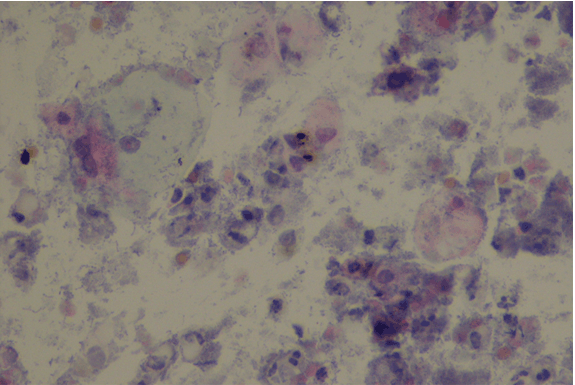
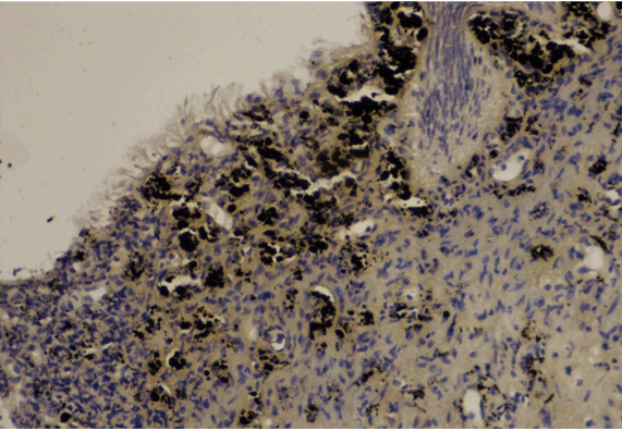
We present the case of a 64-year-old male, former smoker and with a history of previous transurethral resection of prostate with macroscopic hematuria and lower urinary tract symptoms in the last six months. Bladder echography showed generalized thickening of the bladder wall and cytology with abnormal results suggesting repeating. The patient was referred to our institution for further evaluation and under cystoscopy examination, we could observe many clusters of black spots on the bladder wall (Figure 1) and (Figure 2). Ureteric orifices were minor involved and no papillary lesions were identified. Urine cytology was repeated. The first result obtained was bladder cytology that showed reactive transitional cells, with lots of histiocytes, containing a brown-blackish pigment, positive for Masson-Fontana technique (Figure 3). The patient was submitted to a transurethral resection (TUR) of bladder of all macroscopic lesions under general anesthesia because of the clinic suspicion. Bladder biopsy histologically revealed diffuse dark brown-black pigment mostly within the superficial lamina propria, focally in the overlying urothelium (Figure 4). The pigment stained black by Masson-Fontana technique (Figure 5) with flat of punctuated patterns and disappeared after bleaching with potassium permanganate. There was not immunostaining for S100, HMB45 and Melan-A, indicating an absence of mucosal melanocytes. In conclusion, the histopathological result was compatible with primary bladder melanosis. Our patient had a two-year follow-up with annual cystoscopy and cytology, with no signs of recurrence of lesions with normal macroscopic urothelium findings. | ||||||
| ||||||
| ||||||
| ||||||
| ||||||
| ||||||
|
Discussion
| ||||||
|
So far, less than 20 cases have been reported in literature and reviewing current publications we can acknowledge that median patient age is about 60 years (43–86), with no predominance in gender distribution. The presenting symptoms are often nonspecific, and include hematuria (50%), incontinence (17%), recurrent cystitis (11%), dysuria (11%), overactive bladder (11%) and obstructive voiding (5%). On cystoscopy, melanosis of the bladder is characterized by multifocal and diffuse melanin pigmentation of the urothelial mucosa. The bladder is found to be dark brown or black in appearance and deposits can be found at any location in the bladder. This dark color can be due to melanosis, melanoma, hemosiderin or lipochrome deposits and these can be differentiated by histological examination. Histopathological evaluation of bladder biopsies shows aggregates of pigmented granules within urothelial cells and/or similar granules within macrophages within the lamina propria [6]. Histologically, standard H&E staining does not reliably distinguish between melanin and other possible types of dark pigment, but staining of the granules is enhanced by Masson-Fontana stain or Schmorl's reaction, and the pigmentation is abolished by bleach exposure. Pigmentation does not extend deeper than the lamina propria. Usually, Prussian blue and long Ziehl–Neelsen stains are negative, confirming that the pigmented granules are neither hemosiderin nor lipofuscin. These features support the notion that the granules are indeed melanin [3]. Differential diagnosis includes primary or metastatic melanoma, hemosiderin or lipochrome pigment deposition and blue nevus. As for primary or malignant melanoma metastatic to the bladder, can be excluded by careful histopathological examination of the morphologic findings and negative staining for S100, HMB45 and Melan-A [8]. The longest reported follow-up in literature is 10 years, and, as said, many authors believe that bladder melanosis is a benign lesion, but reported cases of associated malignancy, sustained the need for regular cystoscopy and cytology, at least, every year. The need for further evaluation or biopsies should be decided based on clinical evidence. | ||||||
|
Conclusion
| ||||||
|
Bladder melanosis of the urinary bladder is a rare condition, often accompanying with urinary symptoms (half of the cases with reported macroscopic hematuria). Initial evaluation should include cystoscopy and, eventually, upper urinary imaging. Diagnosis is done by endoscopic biopsy and can rule out associated malignant lesions or other melanocytic lesions of the bladder. The possibility that bladder melanosis can be associated with premalignant and malignant urothelial lesions, suggests a close follow-up with, at least, annual cystoscopy and cytology. In our case cytology had a pivotal role in the early diagnosis and characterization of the disease. | ||||||
|
References
| ||||||
| ||||||
|
[HTML Abstract]
[PDF Full Text]
|
|
Author Contributions
Ricardo Godinho – Substantial contributions to conception and design, Acquisition of data, Analysis and interpretation of data, Drafting the article, Revising it critically for important intellectual content, Final approval of the version to be published Edgar Tavares – Substantial contributions to conception and design, Acquisition of data, Analysis and interpretation of data, Drafting the article, Revising it critically for important intellectual content, Final approval of the version to be published Rui Oliveira – Substantial contributions to conception and design, Acquisition of data, Analysis and interpretation of data, Drafting the article, Revising it critically for important intellectual content, Final approval of the version to be published Paulo Conceição – Analysis and interpretation of data, Drafting the article, Revising it critically for important intellectual content, Final approval of the version to be published |
|
Guarantor of submission
The corresponding author is the guarantor of submission. |
|
Source of support
None |
|
Conflict of interest
Authors declare no conflict of interest. |
|
Copyright
© 2016 Ricardo Godinho et al. This article is distributed under the terms of Creative Commons Attribution License which permits unrestricted use, distribution and reproduction in any medium provided the original author(s) and original publisher are properly credited. Please see the copyright policy on the journal website for more information. |
|
|