 |
|
Case Report
| ||||||
| Testicular metastasis from colorectal carcinoma: A case report | ||||||
| Nader Aldossary1, Hend Alshamsi2, Riyad T. Al Mousa3 | ||||||
|
1Urology Resident, Urology Department, King Fahd Specialist Hospital-Dammam, Dammam, Saudi Arabia
2Medical student, Imam Abdulrahman Alfaisal University, Dammam, Saudi Arabia 3Consultant NeuroUrologist, Urology Department, King Fahd Specialist Hospital-Dammam, Dammam, Saudi, Arabia | ||||||
| ||||||
|
[HTML Abstract]
[PDF Full Text]
[Print This Article]
[Similar article in Pumed] [Similar article in Google Scholar] |
| How to cite this article |
| Aldossary N, Alshamsi H, Al Mousa RT. Testicular metastasis from colorectal carcinoma: A case report. J Case Rep Images Urol 2017;2:8–13. |
|
ABSTRACT
| ||||||
|
Introduction: Colorectal cancer is one of the most common cancers worldwide. Metastasis of colorectal cancer to the testis is very rare and only few cases were reported in literature in which a primary colorectal cancer metastasized to the testes. To date, only 42 cases of primary colon cancers metastasizing to testes were reported in literature. | ||||||
|
INTRODUCTION
| ||||||
|
Colorectal cancer is one of the most common cancers worldwide [1]. Colorectal metastasis is considered common especially in patients with recurrence [1]. The most common site for colorectal cancer metastasis is the liver, followed by the lungs, loco-regional, intraabdominal, retroperitoneal and peripheral lymph nodes [1] Metastasis to the testes is considered very rare and only few cases were documented in literature of primary tumor of the colon metastasizing to testis. This is a report of colorectal carcinoma metastasizing to the testis and review of literature. | ||||||
|
CASE REPORT
| ||||||
|
A 47-year-old male was diagnosed with colorectal adenocarcinoma on February 2008. His past medical and surgical histories were unremarkable. Left hemi-colectomy and colostomy were done on March, 2008 and re-anastomosis surgery was done three months later. He received two cycles of chemotherapy but this did not control the disease and metastasis to liver and retroperitoneal lymph nodes on postoperative computed tomography scan of chest, abdomen and pelvis were found. The patient was referred to the urology clinic on April 2009 with painful right testicular swelling of four months’ duration. No lower urinary tract symptoms were found. On physical examination, the patient appeared cachectic. He was vitally stable. Abdominal examination showed ascites with no evidence of tenderness. Genital examination revealed right testicular mass, around 3x2 cm in size, which was slightly tender with positive transillumination test. The left testis was normal. No evidence of scrotal skin changes. phallus is unremarkable. Complete blood count, renal function and coagulation profile were normal with negative urine culture. Tumor markers were done and showed CEA of 512 ng/mL (normal range 0–5 ng/mL), AFB of 2.51 ng/mL (normal range 1.09–8.04 ng/mL), LDH of 157 U/L (normal range 90–215 U/L) and ß-hCG of <1.2 mIU/ml (normal range 0–5 mIU/ml). Scrotal ultrasound was done on November 2008 and showed right testicular hypoechoic mass with irregular outlines (2.1x1.7 cm) infiltrating scrotal wall with hydrocele (4x2.8 cm) (Figure 1). The left testis was normal in size, echogenicity and blood flow. A follow-up scrotal ultrasound on April 2009 showed increase in the size of the right testicular mass (3.8x2 cm) with calcific foci and hyper vascularity and increase in the size of the associated hydrocele. The left testis remained normal. No FNAC was done. Palliative right radical orchiectomy was done through an inguinal approach. Intraoperatively, part of the spermatic cord was excised and scrotal skin was free of invasion. The patient was discharged in a stable condition. One week later he was transferred to Palliative Care and signed do not resuscitate (DNR). Histopathology report revealed a metastatic moderately differentiated adenocarcinoma with mucinous differentiation. The tumor is partially cystic and seen at the spermatic cord resection margin (Figure 2). He was seen in the clinic of urology once after the procedure but unfortunately, he passed away 4 months later due to progression of his primary disease. | ||||||
| ||||||
|
DISCUSSION
| ||||||
|
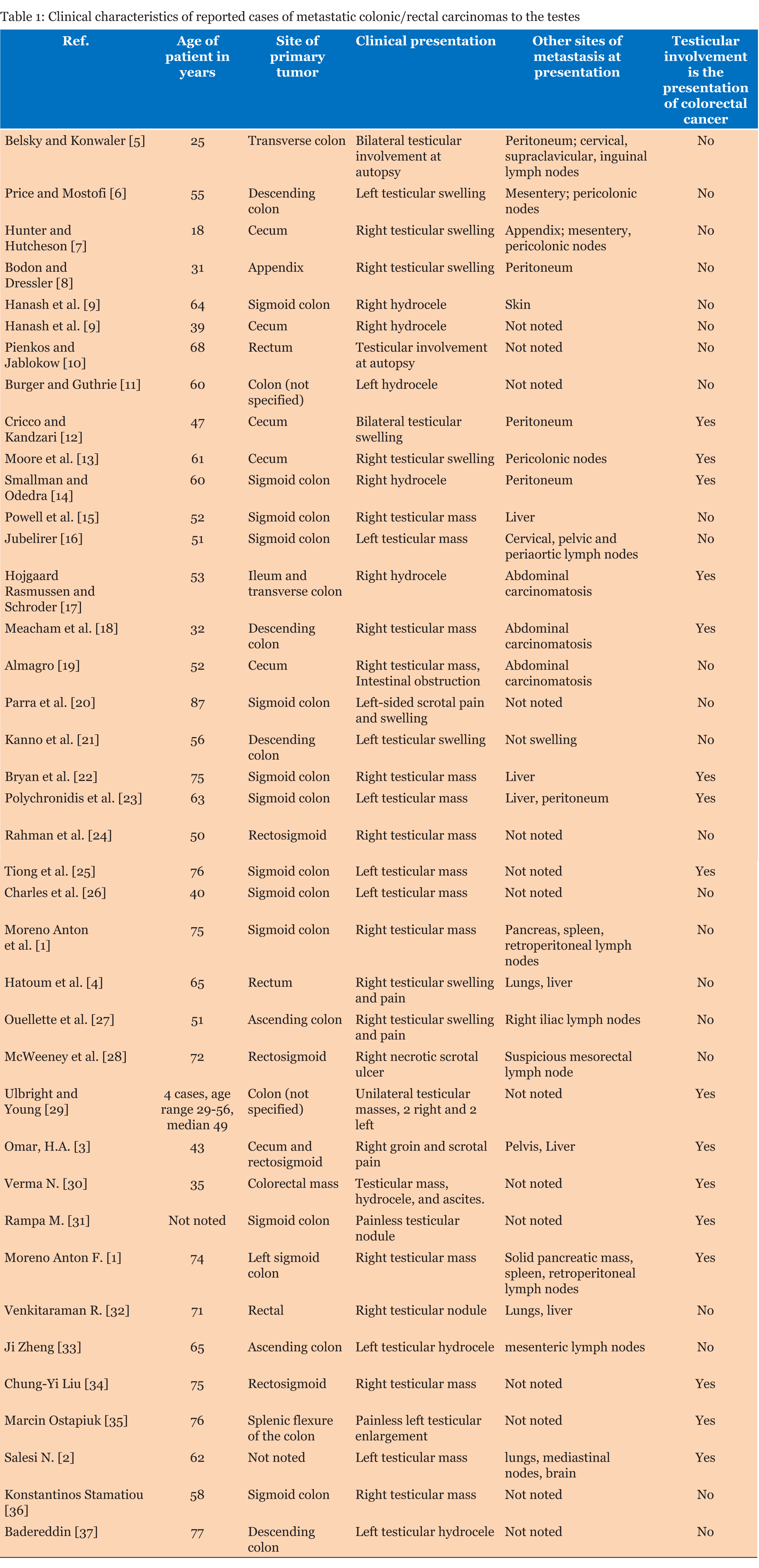
Metastatic carcinoma to the testis is very rare [1]. This is probably due to the unfavorable temperature of the scrotum as metastatic tumor cells find it difficult to survive in such environment as suggested by some researchers [2]. The most common tumors that metastasize to the testis are: prostate (35%), lungs (18%), melanoma (18%), kidney (9%), and colorectal in less than 8% [3]. Since 1988, approximately 35 cases have been documented in the international literature for metastatic carcinoma to the testis [3]. The exact mechanism of spread to the testes is still not fully understood, but few theories have been suggested [4]. These include retroperitoneal lymph nodes/lymph canals involvement, direct involvement which may result in communicating hydrocele, arterial and venous embolization and retrograde sperm duct extension [4]. Most cases of metastases to the testes were discovered accidently by autopsy, and only a few cases presented with communicating hydrocele and rarely as a solid tumor in testis [3]. In our case, the patient presented with both a communicating hydrocele and a solid mass of testis. It is important to differentiate primary from secondary tumors of testis [4]. The differentiation is done mostly by histopathological evaluation of the resected tumor. Other differentiating factors are age at diagnosis, past history of any tumor, and imaging studies [4]. Determining the origin of tumor is nowadays done by immunohistochemistry (IHC); for example in colorectal metastasis to testis, the CEA and cytokine 20 (CK-20) staining are positive [4]. Early suspicion and diagnosis of such case allows palliative surgical intervention, which leads to relieving the patients’ symptoms, controlling tumor growth and nowadays improving the survival of the patient. In this case, the patient underwent surgery of the colon and post-operative chemotherapy which failed to control the disease. The patient developed metastasis to liver and retroperitoneal lymph nodes, and after one year he developed metastasis to the testis. A previous literature review, from 1950 through January 2010, yielded 33 cases of testicular metastasis from rectal or colonic carcinoma. Clinical information was not available for 2 of the 33 reported cases [4]. We reviewed literature further and added nine more cases from 2010–2017 making the total reported cases of testicular metastases from colorectal carcinoma 42 cases as given in Table 1 [1][2][3] [4][5][6] [7] [8][9][10][11][12][13][14][15][16][17][18][19][20][21][22][23][24][25][26][27][28][29][30][31][32][33][34][35][36][37]. | ||||||
| ||||||
|
CONCLUSION
| ||||||
|
Metastatic colorectal carcinoma to urogenital tract is very rare and because of this the patients usually present late with advanced unresectable tumor. This must be kept in mind and physicians caring for these patients should be aware of this. A patient with history of previous colorectal carcinoma and similar presentation should raise suspicion of metastatic colorectal carcinoma to the testis. Surgery should be considered as palliative to relieve the patients’ symptoms, control the tumor growth, and improve the survival outcome. | ||||||
|
REFERENCES
| ||||||
| ||||||
|
[HTML Abstract]
[PDF Full Text]
|
|
Author Contributions
Nader Aldossary – Substantial contributions to conception and design, Acquisition of data, Drafting the article, Final approval of the version to be published Hend Alshamsi – Substantial contributions to conception and design, Acquisition of data, Drafting the article, Final approval of the version to be published Riyad T. Al Mousa – Substantial contributions to conception and design, Analysis and interpretation of data, Revising it critically for important intellectual content, Final approval of the version to be published |
|
Guarantor of Submission
The corresponding author is the guarantor of submission. |
|
Source of Support
None |
|
Conflict of Interest
Authors declare no conflict of interest. |
|
Copyright
© 2017 Nader Aldossary et al. This article is distributed under the terms of Creative Commons Attribution License which permits unrestricted use, distribution and reproduction in any medium provided the original author(s) and original publisher are properly credited. Please see the copyright policy on the journal website for more information. |
|
|


